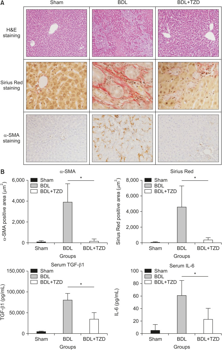
Fig. 1. Liver fibrosis following BDL was attenuated in BDL+TZD rats. (A) Representative stained liver sections in sham, BDL, and BDL+TZD groups, stained using H&E (original magnification, ×10) and fibrosis assessment using Sirius Red staining (original magnification, ×40) and α-SMA staining (original magnification, ×20). (B) α-SMA-positive immunostaining area, Sirius Red-positive staining area, and serum levels of TGF-β1 and IL-6 levels in sham, BDL, and BDL+TZD rat groups; all variables are significantly higher in BDL than BDL+TZD groups.
BDL: bile duct ligation, TZD: thiazolidinedione, α-SMA: α-smooth muscle actin, TGF-β1: transforming growth factor beta1, IL-6: interleukin-6. *p<0.05.

