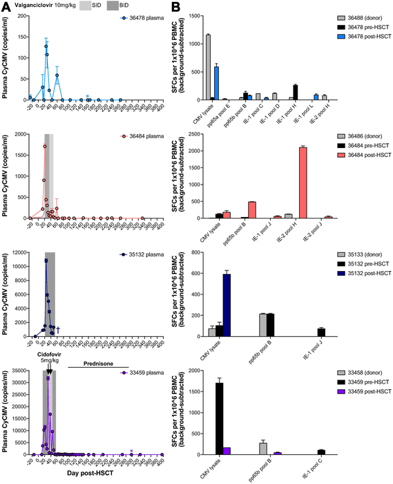
Figure 4. Cytomegalovirus consistently reactivates early post-transplant, but is subsequently controlled.
A, Longitudinal cynomolgus Cytomegalovirus (CyCMV) plasma viral loads in four representative HSCT recipient macaques, measured by quantitative PCR (limit of detection 60 DNA copies/ml). Gray boxes indicate periods of oral valganciclovir treatment. Arrows indicate intravenous cidofovir treatment. Colored asterisks indicate timepoints at which post-HSCT PBMC was tested for CMV-specific T cell responses, shown in part B. Dark blue cross indicates timepoint of 35132 euthanasia. SID = once daily, BID = twice daily. B, CMV-specific T cell responses measured by interferon-gamma ELISPOT of peripheral blood mononuclear cells (PBMC) stimulated with CMV lysate or pools of 15-mer peptides spanning IE-1, IE-2, and pp65 (11 amino acid overlap). Graphs show results for HSCT recipient macaques shown part A, pre-HSCT (black) and post-HSCT (colored), and their HSCT donors (gray). SFCs = spot-forming units.

