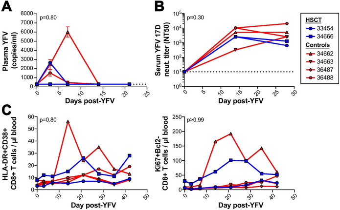
Figure 5. Long-term engrafted HSCT recipient macaques respond to live-attenuated yellow fever 17D vaccination similarly to untransplanted macaques.
A, Longitudinal yellow fever (YFV) plasma viral loads in two long-term engrafted HSCT recipient macaques (blue) compared to four control untransplanted Mauritian cynomolgus macaques (red) after live-attenuated yellow fever 17D vaccination. Undetectable plasma viral loads graphed as 300 RNA copies/ml, the limit of detection (LOD) for the assay, indicated by horizontal dotted line. B, Longitudinal serum YFV 17D neutralizing antibody titers. NT50 = highest fold serum dilution where YFV 17D plaque number was reduced by at least 50%. The lowest dilution of serum tested was 10-fold, indicated by the horizontal dotted line. C, Longitudinal counts of activated CD8+ T-cells in blood post-vaccination, as measured by HLA-DR and CD38 (left) and Ki67 and Bcl2 (right). No statistically significant difference in plasma viral loads or CD8+ T cell activation were observed between the HSCT and control groups, analyzed by comparing areas under the curve (AUC) using the Mann-Whitney test (p-value displayed on graphs). No statistically significant difference in neutralizing antibody titers were observed between the HSCT and control groups at any timepoint, analyzed using repeated measures ANOVA (overall group factor p-value displayed on graph, Sidak-adjusted p-values for timepoints 0, 14, and 28 are >0.99, 0.76, and 0.37, respectively).

