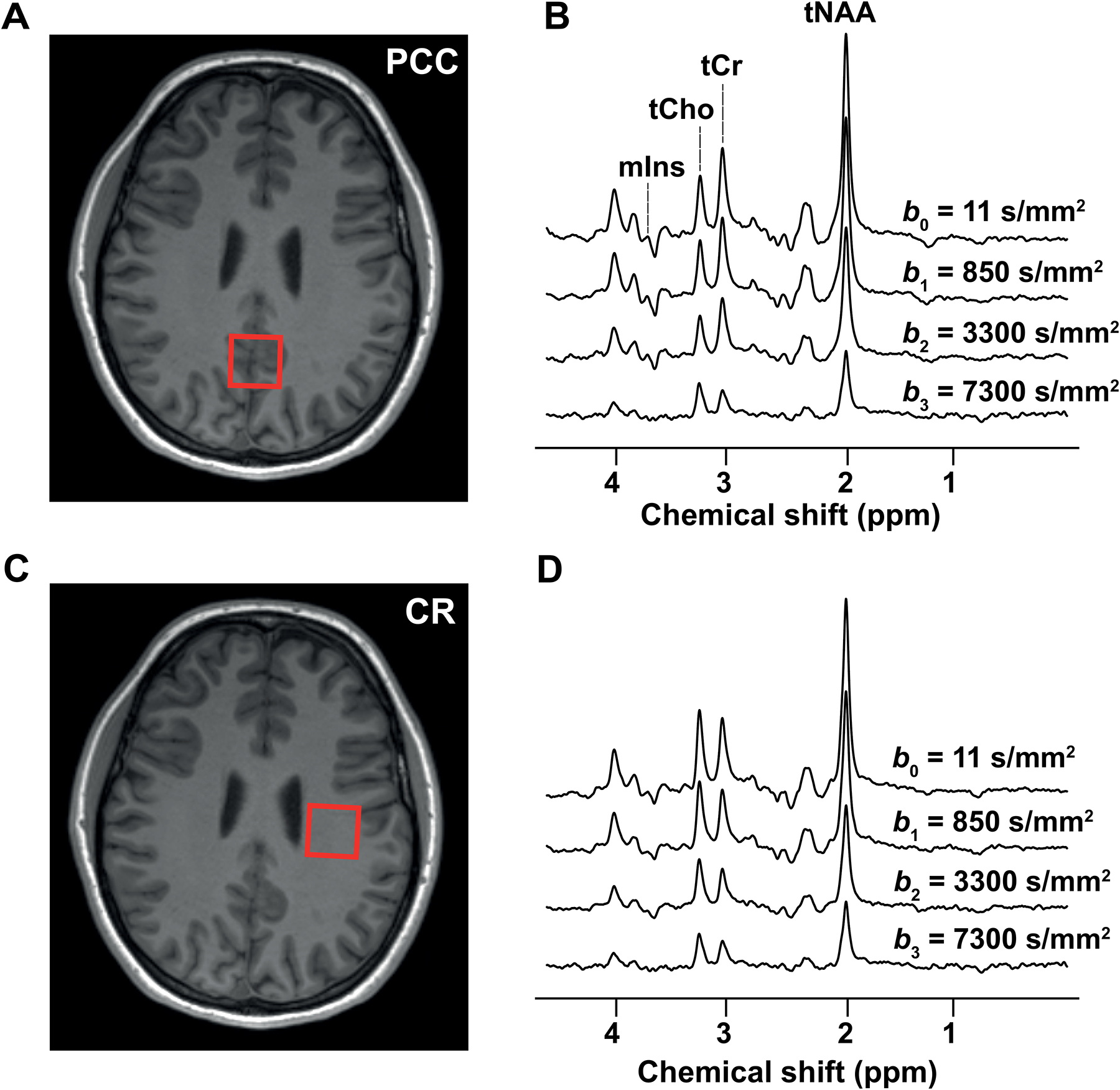
Figure 2: Diffusion-weighted spectra and VOIs.

The locations of the VOIs in (A) the PCC and (C) the CR are shown on T1-weighted images together with examples of diffusion-weighted spectra acquired at different b-values in (B) VOIPCC and (D) VOICR

The locations of the VOIs in (A) the PCC and (C) the CR are shown on T1-weighted images together with examples of diffusion-weighted spectra acquired at different b-values in (B) VOIPCC and (D) VOICR