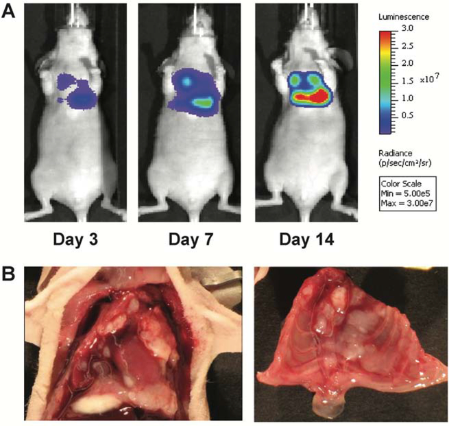
Figure 1. Murine orthotopic xenograft model of malignant pleural mesothelioma.
(A) After intrathoracic (IT) injection of 106 MSTO-211H-luciferase cells, animals were serially imaged on days 3, 7, and 14. Representative images show that tumor is clearly established in the chest at day 3 with progression to significant tumor burden over time. (B) Representative necropsy findings show multiple pleural tumor deposits diffusely distributed throughout the bilateral chest cavities.

