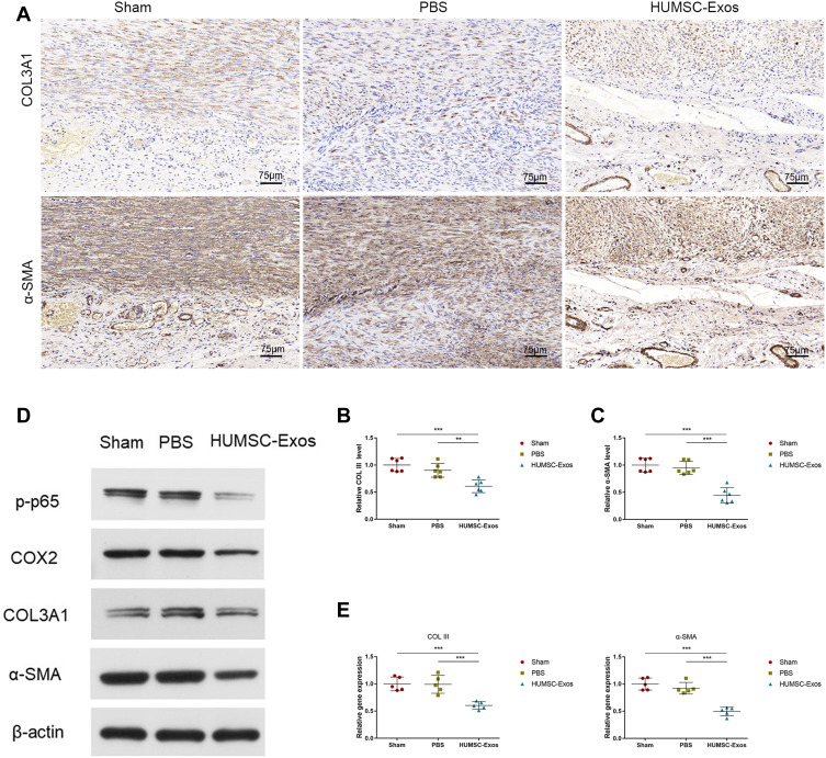
Figure 4.
HUMSC-Exos synchronously inhibited rat tendon fibrosis and p65 activity. (A) Immunohistochemical staining of COL III and α-SMA. Scale bar: 75 μm. (B and C) Quantitative analysis of COL III and α-SMA (n = 6 per group). (D) WB images of COL III and α-SMA. (E) Quantitative analysis of mRNA for COL III and α-SMA (n = 5 per group). **P < 0.01, ***P < 0.001.

