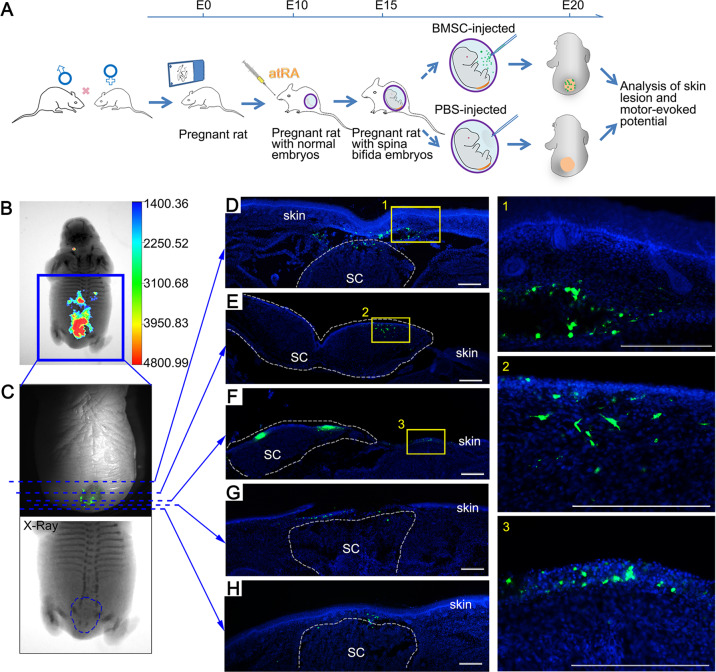
Fig. 5. Transplanted BMSCs engrafted in different tissues of the lesion.
a Experimental design and time course: intragastric atRA administration was performed on the morning of E10, the intrauterine BMSC microinjection was performed on E15, and analysis of skin lesions and motor-evoked potential (MEPs) of the fetuses were performed after cesarean section on E20. b Representative overlay images of X-ray and fluorescence images show a large number of GFP+ BMSCs at the defective lumbosacral region. c The fluorescence (top) and X-ray (bottom) images were enlarged from the blue box in A. The blue dotted lines indicate where the fluorescence sections (d–h) were obtained and the blue dotted circle indicates the abnormal lumbar sacrum in the fetus. d–h Representative sections of BMSC engraftment from upper edge of the lesion to the lower edge. The GFP+ BMSCs (green) scattered on the subcutaneous tissue of the defect (d and h), dorsal view of the defective spinal cord (e and f), abnormal skin surface (f), and the place where the skin tissue should be extended to cover the defect (g). Highlighted regions in d–f were magnified and shown on the right. Scale bars: 100 μm.

