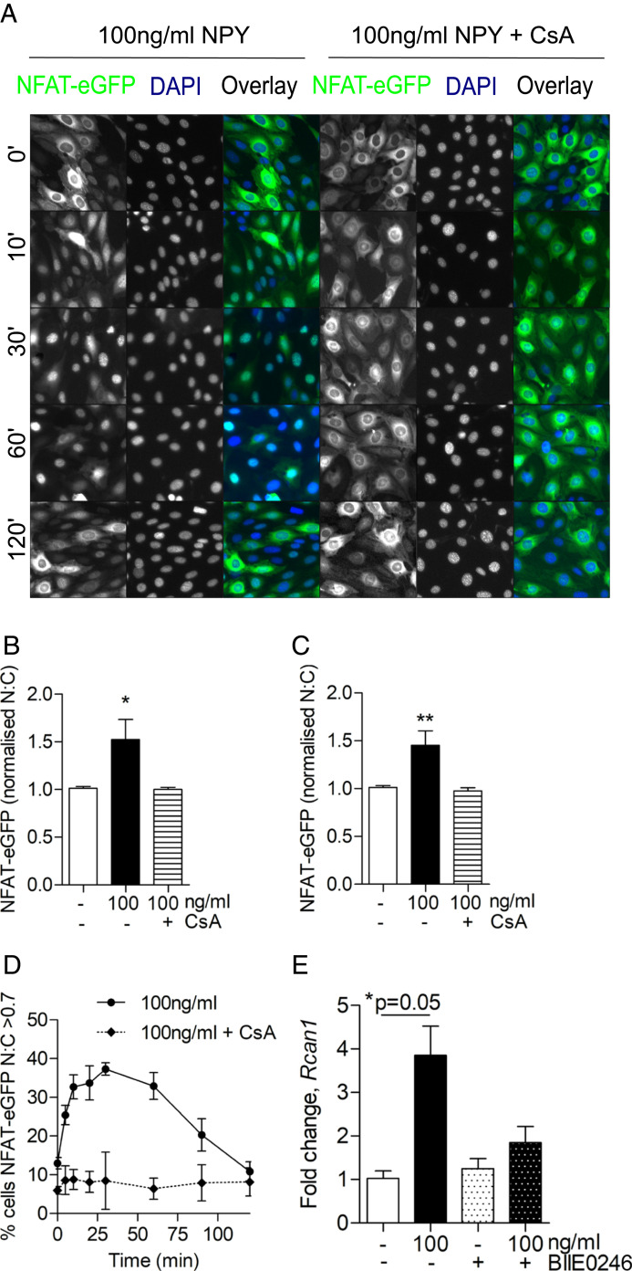
Fig. 7.
NPY stimulates calcium-dependent NFAT activation in podocytes. Podocytes were transfected with NFAT-eGFP and stimulated with 100 ng/mL of NPY at indicated time points before fixation and DAPI staining. Where indicated, 10 μM CsA was added 15 min prior to NPY stimulation. Modest changes in brightness and contrast were applied to all images for visual purposes; unmodified images were used for quantification. (A) Representative fluorescent images. (B and C) Population average values for NFAT-eGFP nuclear:cytoplasmic (N:C) ratios derived from arbitrary fluorescence unit measurements of NFAT-eGFP intensity in nuclear and cytoplasmic compartments and normalized to control (time 0), demonstrating a significant increase in N:C NFAT-eGFP at (B) 30 min and (C) 60 min post stimulation. *P < 0.05 and **P < 0.01, Mann–Whitney U test; no significant differences were observed with CsA pretreatment; n = 3; each condition was tested in triplicate. (D) Percentage of activated cells (where N:C of individual cells >0.7) in whole-cell populations over time; n = 3. (E) qPCR results demonstrating fold change in regulator of calcineurin 1 (Rcan1) expression following NPY stimulation, with and without costimulation of the Y2 receptor antagonist, BIIE0246, at 1 μM for 24 h; *P = 0.05, Mann–Whitney U test; n = 3.

