Fig. 2.
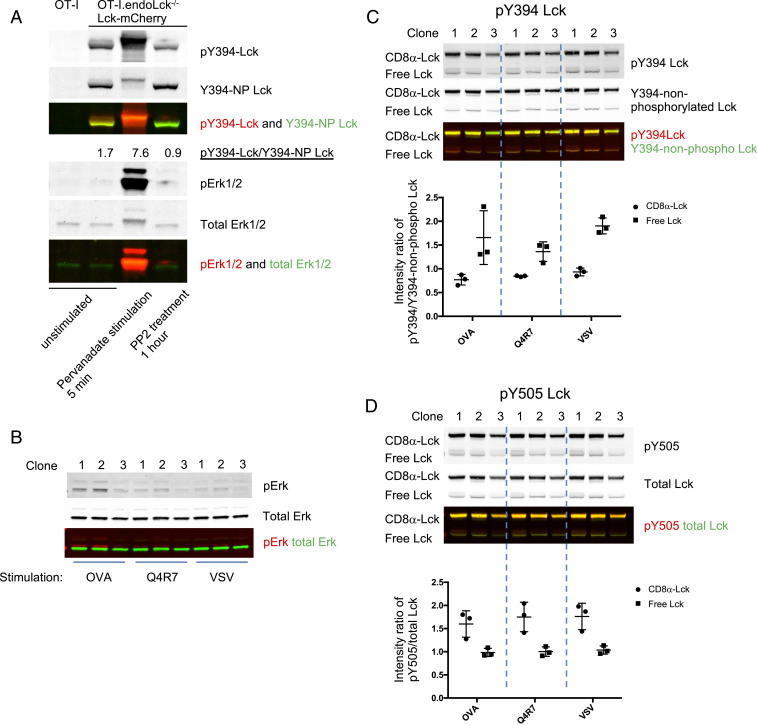
Higher Y394 phosphorylation in free Lck than CD8-bound Lck fraction in T hybridoma cells. (A) OT-I endoLck−/− CD8αβ-expressing hybridoma cells (OT-I.CD8αβ+.endoLck−/−) overexpressing Lck-mCherry were unstimulated, stimulated with Pervanadate (PV) for 5 min, or treated with PP2 inhibitor for 1 h before lysis. Signal intensity ratios of pY394/Y394-NP Lck were calculated. (B–D) OT-I.CD8αβ+.endoLck−/− hybridoma cells transduced with both CD8α-Lck-Cerulean and Lck(C20.23A)-mCherry were sorted as single clones (OT-I.CD8αβ+.endoLck−/−0.8αLckC.LckC2023A.R). The cell clones were stimulated by H2-Kb-OVA/Q4R7/VSV tetramers for 5 min before lysis. The Erk phosphorylation (B), pY394/Y394-NP Lck (C), or pY505/total Lck (D) levels of each cell lysis were detected by WB. The intensity ratio of pY394/Y394-NP Lck or pY505/total Lck of free and coreceptor-bound Lck were summarized. Mean ± SEM is shown. The figure is representative of two independent experiments.

