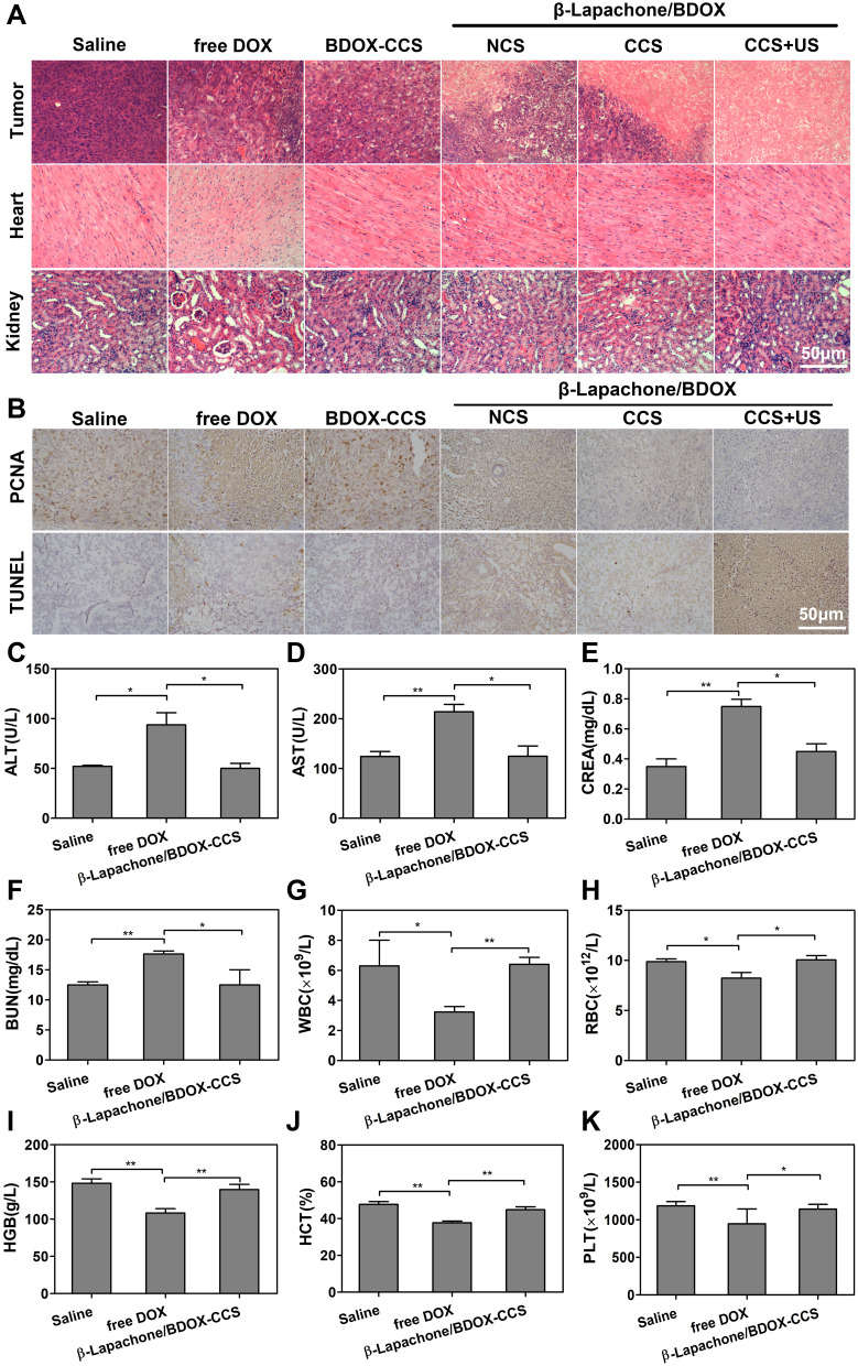
Figure 8.
Pathological analysis in xenograft nude mice with HepG2 tumor. (A) Histological sections of tumor tissues and main organs stained with H&E. (B) PCNA and TUNEL staining of tumors isolated on day 25 for observing proliferation and apoptosis. (C–K) Biochemical studies, including liver functions (ALT, AST), renal functions (CREA, BUN), and hematology data (WBC, RBC, HGB, HCT, PLT) in healthy BALB/c mice treated with saline, free DOX, and β-Lapachone/BDOX-CCS. Data are represented as mean ± SD, n = 3; *p < 0.05, **p < 0.01.

