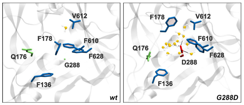
Figure 6.
Close-up of the residue 288 and surrounding residues of the HP trap in STmAcrBWT and STmAcrBG288D. Waters belonging to the first and second hydration shell of residue 288 (distance threshold: 5 Å, see Materials and Methods) are also shown, and hydrogen bonds involving residue 288 are represented as dashed lines. This image has been created using two representative frames of MD trajectories.

