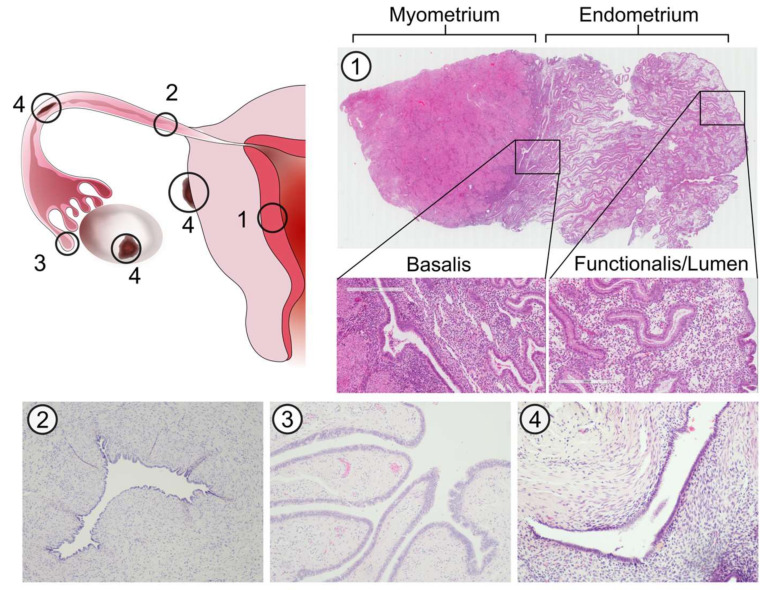
Figure 1.
Histological architecture of the endometrium, fallopian tubes (FT) and endometriotic lesions. Micrographs show H&E stained sections of the endometrium (1), transverse planes of the FT isthmus (2) and fimbriae (3) and an endometriotic lesion (4). The endometrium can be divided into three discrete layers based on epithelial cell phenotype; the basalis, functionalis and lumen. Unlike the endometrium, the FT has a single convoluted layer of epithelial cells lining the lumen (endosalpinx) surrounded by stroma. Endometriotic lesions are characterised by the presence of both stromal and epithelial components, the latter of which may exhibit a glandular or luminal phenotype.

