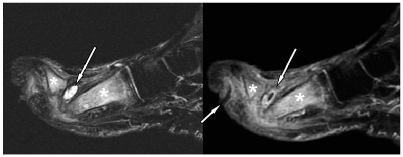
Figure 3.
Forefoot ulcer, sinus track, and abscess associated with OM in a 57-year-old diabetic man with a 16-year history of insulin-dependent diabetes. Sagittal T2 fat-suppressed (left panel), and post-contrast T1-weighted fat-suppressed MRI (right panel) shows a dorsal thick rim-enhancing abscess adjacent to the first metatarsal head (arrows). Note a plantar ulcer appearing as a focal skin interruption and a sinus tract with rim-like enhancement (small arrow in right panel) extending near the first proximal interphalangeal joint. Given these findings, the hyperintensity (** in left panel), and post-contrast enhancement (** in right panel) in the first metatarsal, and proximal phalanx respectively, are indicative of OM.

