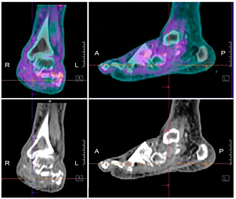
Figure 5.
Example of [18F]FDG PET/CT in a patient with Charcot osteoarthropathy. Fused images (upper panels) show moderate and diffuse uptake, which is interesting in particular bones and joints of the mid and hind-foot. Co-registered low-dose CT images (lower panels) show the evident destruction of bony architecture. These findings are consistent with the diagnosis of Charcot foot, but they do not allow discriminating a pan-inflammation from a possible superimposed infection.

