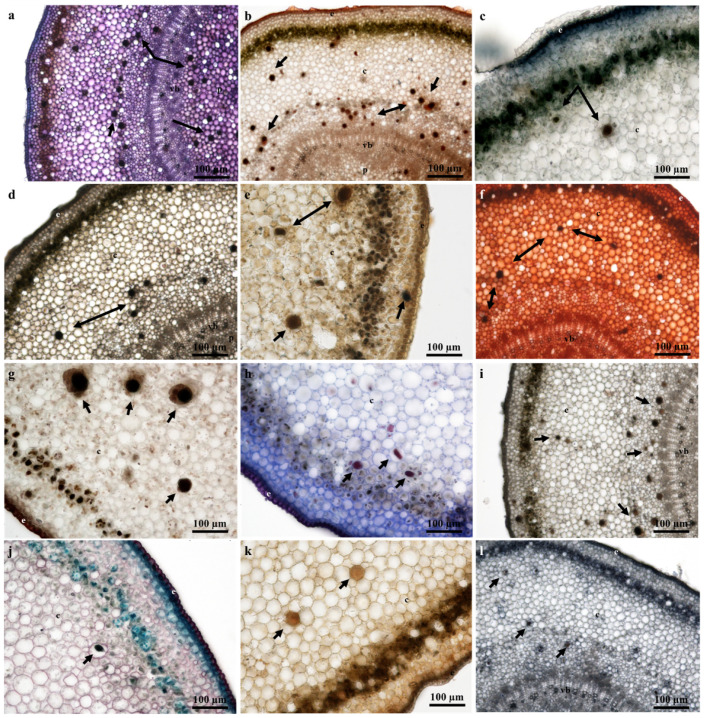
Figure 8.
Histochemical observations of laticifers in young stem sections of T. ventricosa. (a) Presence of polysaccharides in laticifers stained using Toluidine Blue. (b) Lipids stained using Sudan IV. (c) Negative staining of lipids using Sudan Black B. (d) Ferric Trichloride positively stained laticifers a dark-black color. (e) Alkaloids identified within laticifers using Wagner’s and Dittmar’s reagent. (f) Detection of mucilage and pectin using Ruthenium Red. (g) Intense staining of resin acids in laticifers using NADI reagent. (h) Neutral lipids in laticifer identified using Nile Blue. (i) Detection of phenolics within laticifer stained using Ferric Chloride. (j) Presence of acidic substances within laticifers stained using Safranin and Fast Green. (k) Negative staining of lignin aldehydes using Phloroglucinol. (l) Proteins detected in laticifers. Arrows refer to laticifer.

