Figure 4.
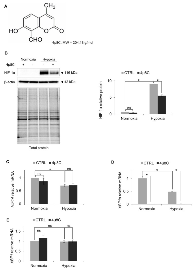
IRE1α inhibition by 4µ8C in hypoxia results in HIF-1α protein reduction in HUVECs. (A) Structural formula of 4µ8C. (B) HIF-1α protein levels after IRE1α inhibition by 4µ8C in normoxia and hypoxia were evaluated by Western Blotting, normalized to β-actin and total protein levels and related to hypoxia (CTRL). The densitometry analysis is representing two independent experiments (* p < 0.05 was considered significant). (C) HIF1A, (D) XBP1s (spliced) and (E) XBP1 (total) mRNA levels were quantified by quantitative real-time PCR, normalized to 18S and RPLP0 rRNA levels and expressed as a fold change over normoxic samples. Data represent the mean ± SEM of two independent experiments.

