Figure 6.
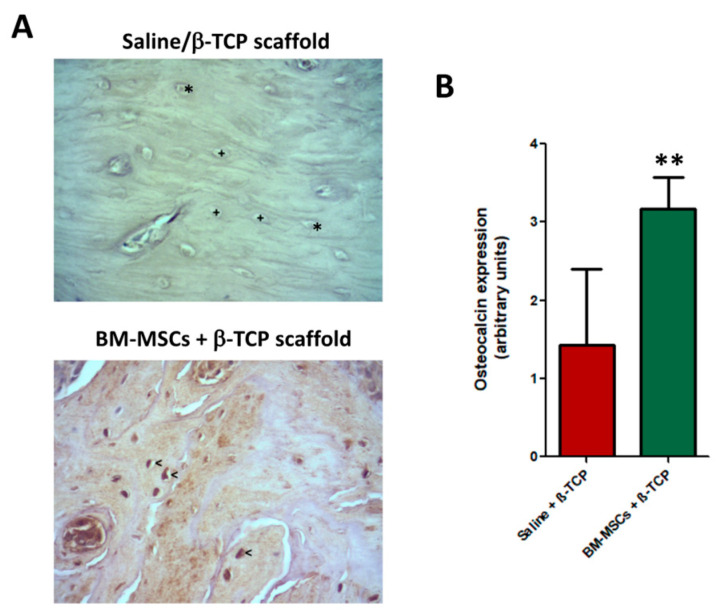
Representative images of immunohistochemical expression of osteocalcin of maxillary bone after induction of osteonecrosis in control animals transplanted only with saline + β-TCP scaffold, (A, top image) and those treated with BM-MSCs + β-TCP scaffold (A, bottom image) 4 weeks after treatment. While in control animals the empty lacunae (+) and osteocytes (*) are negative, osteocytes from BM-MSC-treated bone strongly expressed osteocalcin (<). ABC immunohistochemical procedure anti-osteocalcin were performed. Magnification: ×400; (B) number of osteocalcin positive cells were significantly more abundant, ** p < 0.01.

