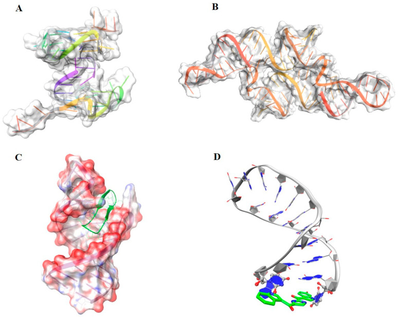
Figure 3.
Example of nucleic acid targets. (A) Structure of the G-quadruplex oncogene c-Myc shown with transparent surface and backbone as cartoon style (PDB code: 6AU4). (B) Example of riboswitch from Glutamine II Riboswitch (PDB code: 6QN3), represented with transparent surface and the backbone as cartoon style. (C) Representation of the HIV trans-activation response (TAR) RNA bound to a high affinity macrocycle (PDB code: 6D2U). The electrostatic surface highlights the charge distribution, whereas the ligand bound is shown in licorice (green). (D) Structure of TSL2 hairpin bound with the small molecule splicing modulator PK4C9 (green). The RNA structure is shown in grey cartoon style and the ligand as licorice [47].

