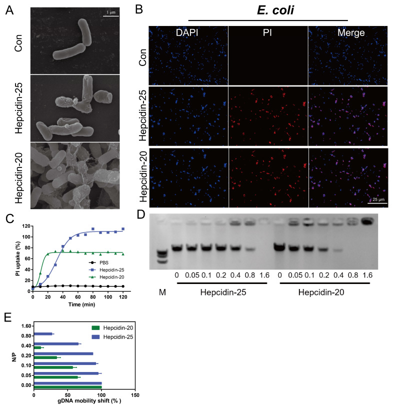
Figure 5.
Sterilization mechanism of hepcidin-20 and -25. (A) Scanning electron microscopy of E. coli cultured with or without hepcidin-20/25 (16 μM) for 2 h at 37 °C. (B) PI update by E. coli after incubated with 16 μM hepcidin-20 or hepcidin-25 for 2 h at 37 °C. Bacteria were stained with DAPI (blue) or PI (red) and imaged using a confocal microscope. (C) Using the PBS as the control, PI uptake of E. coli by hepcidin-20 (green) and -25 (blue). (D) Gel shift assay of bacterial gDNA mixed with increasing concentrations of hepcidin-25 (left) and -20 (right). The abscissa represents the N/P molar ratio (amino nitrogen (NH3+) of peptides/phosphate (PO4−) of DNA). (E) ImageJ was used to analyze the intensity of nucleic acid bands in gel shift assay. The abscissa represents the N/P molar ratio and the ordinate represents the gDNA.

