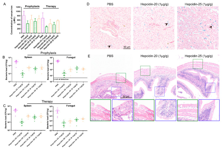
Figure 9.
The effect of hepcidin on iron homeostasis and bacterial loads of spleen and foregut infected with A. hydrophila. (A) The serum iron assay kit quantified the iron content in serum. (B,C) At 3 days post-infection, bacterial loads in spleen and foregut of grass carp infected with A. hydrophila in prophylaxis (B) and therapy (C) groups were detected, (n = 5). (D) At 3 days post-infection, using the PBS group as the control, the iron content in hepatopancreas was stained by Prussian blue, bar = 50 μm. Arrowheads in the image show the iron content in hepatopancreas. (E) At 3 days post-infection, the intestinal tissue sections in the 1 μg/g hepcidin-20 and -25 group were observed, serous membrane (green) and intestinal villi (blue).

