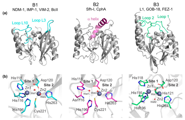
Figure 2.
Representative structures of the three metallo-β-lactamases (MBLs) subclasses: B1 NDM-1 (PDB 3spu), B2 Sfh-I (PDB 3sd9), and B3 L1 (PDB 1sml). (a) Overall protein structures. Loops and helices involved in substrate interactions are shown in color. (b) Active sites. Zn(II) ions are shown as grey spheres and water/OH molecules as red spheres. Zn(II) interactions are shown as dashed lines and metal ligands are shown in color. Residues are numbered according to the standard MBL numbering scheme. For the B2 enzyme Sfh-I, the active mono-Zn(II) form is shown.

