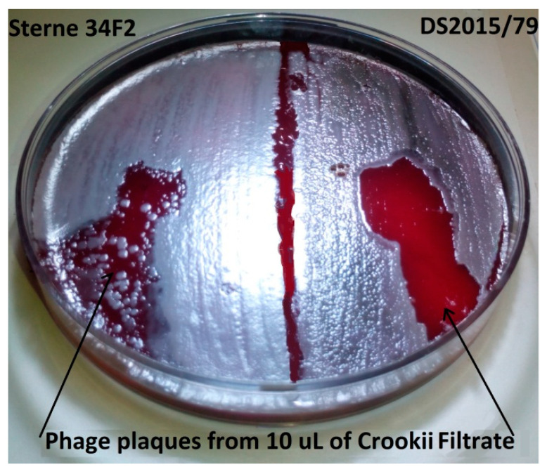
Figure 1.
Blood agar plate with Bacillus anthracis bacterial lawns of Sterne 34F2 (left) and virulent DS201579 (right) after 12 h incubation at 37 °C. The plaques indicated by the arrows are where the bacteriophage Bacillus phage Crookii has lysed bacterial cells. The phage demonstrates a greater affinity for B. anthracis DS201579 than Sterne strain; where the DS201579 lawn is also displaying turbidity indicating phage infection.

