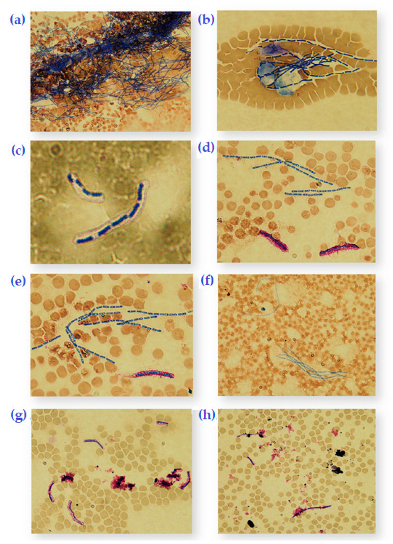
Figure 2.
Romanowski-Giemsa-stained blood smears of Bacillus anthracis at 1000x magnification. (a) Unencapsulated B. anthracis Sterne vaccine strain at 12 h of 37 °C incubation. (b) Sterne with γ-phage at 8 h of 37 °C incubation. (c) B. anthracis DS201579 showing encapsulation (balloon-like layer) at 8 h of 37 °C incubation. (d) DS201579 with NaHCO3 in 8% CO2 at 8 h of 37 °C incubation showing thickened capsule (purple) and cells with endospores (blue). (e) DS201579 at 37 °C incubation for 12 h in 8% CO2 where all cells contain endospores (blue) and degradation of mother cell has begun (purple streaks). (f) DS201579 with NaHCO3 and 8% CO2 at 37 °C incubation for 12 h of showing 100% sporulation. (g) DS201579 with Bacillus phage Crookii at 37 °C incubation for 12 h and 8% CO2 of showing lysed cells as well as intact endospores. (h) DS201579 with Bacillus phage Crookii at 37 °C incubation for 12 h where all vegetative cells in view were lysed.

