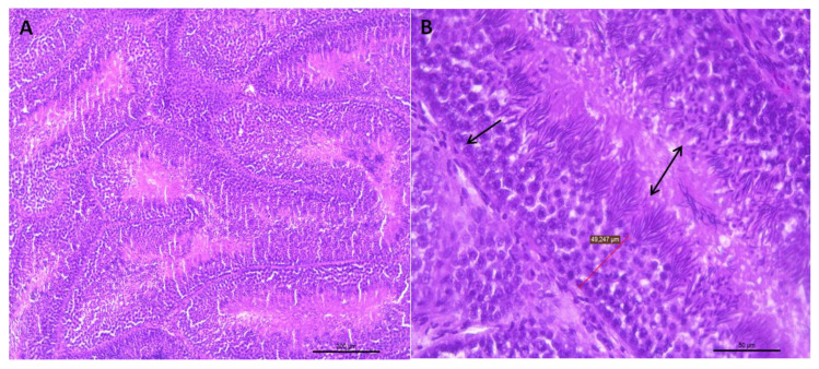
Figure 1.
(A) Representative transverse section of seminiferous tubules of the testes of roosters from 30 weeks of age with standard weight and a value of 3 for testicular integrity. Small dark bodies are observed that correspond to different stages of sperm maturation. HE staining (bar: 200 μm). (B) Area extension of photograph A, where the epithelium of the seminiferous tubule is shown (arrow). This epithelium is formed by germ cells, which, as they move towards the tubule lumen, can be found in different spermatogenesis stages. Towards the tubule lumen, mature sperm clusters can be seen in which the head and the tail can be differentiated (double-headed arrow). HE staining (bar: 50 μm).

