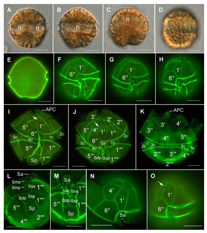
Figure 5.
Light micrographs of Alexandrium pacificum using differential interference contrast (A–D) and epifluorescence microscopy (Solophenyl Flavine staining) of Lugol-fixed cultured cells (E–O). (A) Ventral view with focus in the cell middle, notice the two ends of the sausage-shaped nucleus (n). (B) Dorsal view with focus in the dorsal half of the cell showing the path of the nucleus (n). (C) Antapical view with focus in the cell middle, notice the sausage-shaped nucleus (n) dorsally. (D) Dorsal view with focus on the peripherally located, elongated chloroplasts. (E) General cell shape. (F–H) Ventral view showing the shapes of the first apical (1′) and sixth precingular (6′′) plates. Note the different degree of asymmetry in 1′. (I,J) Ventral views of squeezed thecae showing the thecal plates. (K) Doral view of a squeezed theca showing the thecal plates. (L) Ventral to antapical view showing sulcal plates. (M) Sulcal plates. (N) Ventral epithecal view showing the shapes of characteristic plates 1′, 6′′, and anterior sulcal plate (Sa). (O) The first apical plate (1′) with ventral pore (arrow). Scale bars = 10 µm.

