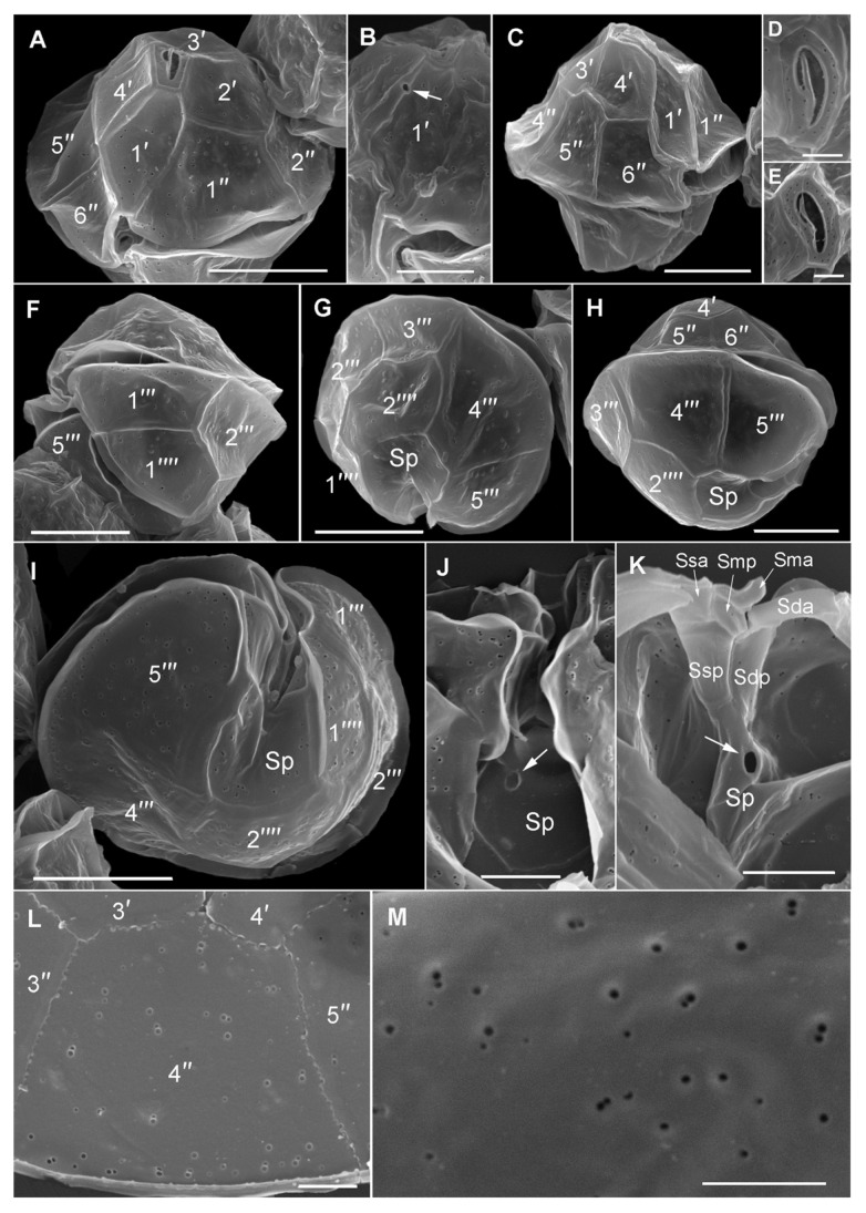
Figure 6.
Scanning electron microscopy of Alexandrium pacificum. (A) Ventral of the epitheca. (B) Detail of a first apical plate (1′) with ventral pore (arrow). (C) Right lateral to ventral cell view. (D,E) Apical pore complex. (F) Left lateral view of a hypotheca. (G) Hypotheca in antapical view. (H) Right lateral cell view. (I) Hypotheca in antapical view showing the sulcus. (J) Detail of the posterior sulcal plate (Sp) with posterior connecting pore (arrow). (K) Inside view of the sulcal area of the hypotheca showing sulcal plate details. Note the posterior connecting pore (arrow). (L,M) Thecal pore pattern and sizes. Scale bars = 10 µm, (B,J,K) 5 µm, (D,E,L,M) 2 µm.

