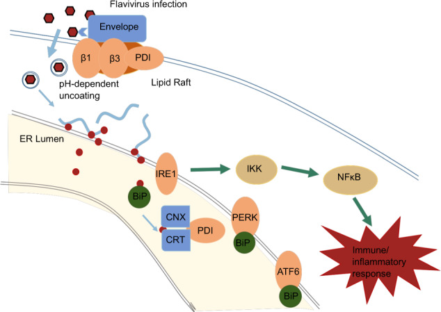
Fig. 7.

An example of PDI function and ER stress response during Flavivirus infection. Flavivirus entry into endothelial cells could be suppressed by silencing of PDI. Studies also show that PDI co-localizes with cell surface lipid rafts along with flavivirus envelope protein, leading to the activation of cell surface integrins (b1 and b3), which has direct implications in aiding the virus entering host cells. Viral RNA is then released and located around ER for translation. The increased protein synthesis may disturb ER homeostasis, leading to unfolded protein response through GRp78/BiP dissociation with PERK and ATF6 to activate PERK, ATF6, and IRE1. Downstream responses include activation of NF-κB and other signal pathways, followed by immune and inflammatory responses
