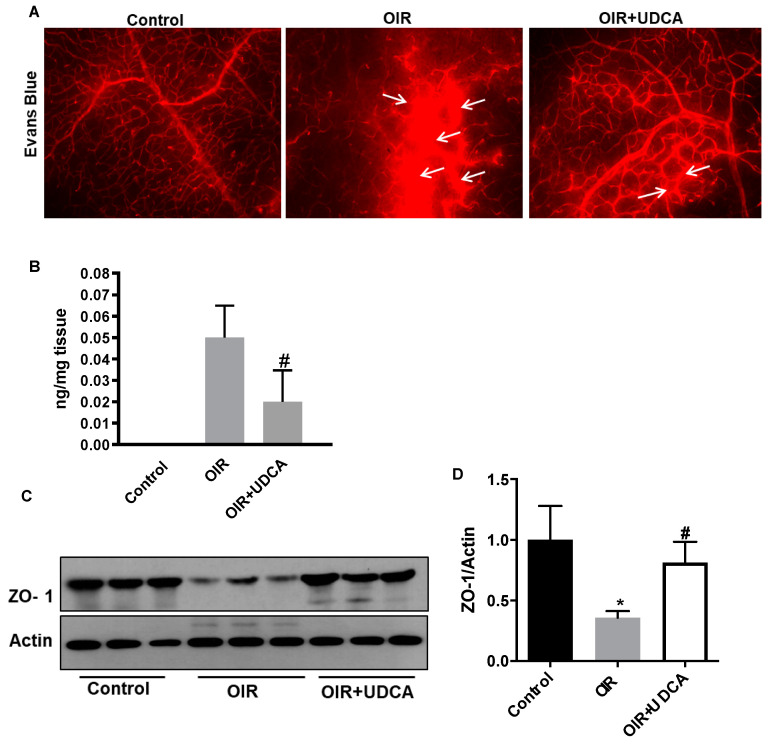
Figure 4.
UDCA preserves blood–retinal barrier in OIR mice. (A) Evans blue (EB) dye extravasation (white arrows) assessed at P17, in control mice (room air), OIR mice, and OIR+UDCA mice (treated with 50 mg/kg UDCA from P7-P17). Two hours after EB injection, eyes were collected to prepare flat mounts and examined using a fluorescence microscope. (B) In a separate set of experiments, EB-injected mice were perfused with phosphate-buffered saline (PBS), and the residual dye in the retina was quantified following formamide extraction. (C) Representative image of zonula occludin-1 (ZO-1) immunoblotting from the different treatment groups. (D) Densitometry results showing changes in ZO-1 protein expression normalized to the loading control actin. Values are mean ± S.D. (n = 6 retinas per group). * p < 0.05 vs. control and # p < 0.05 vs. OIR.

