Figure 5.
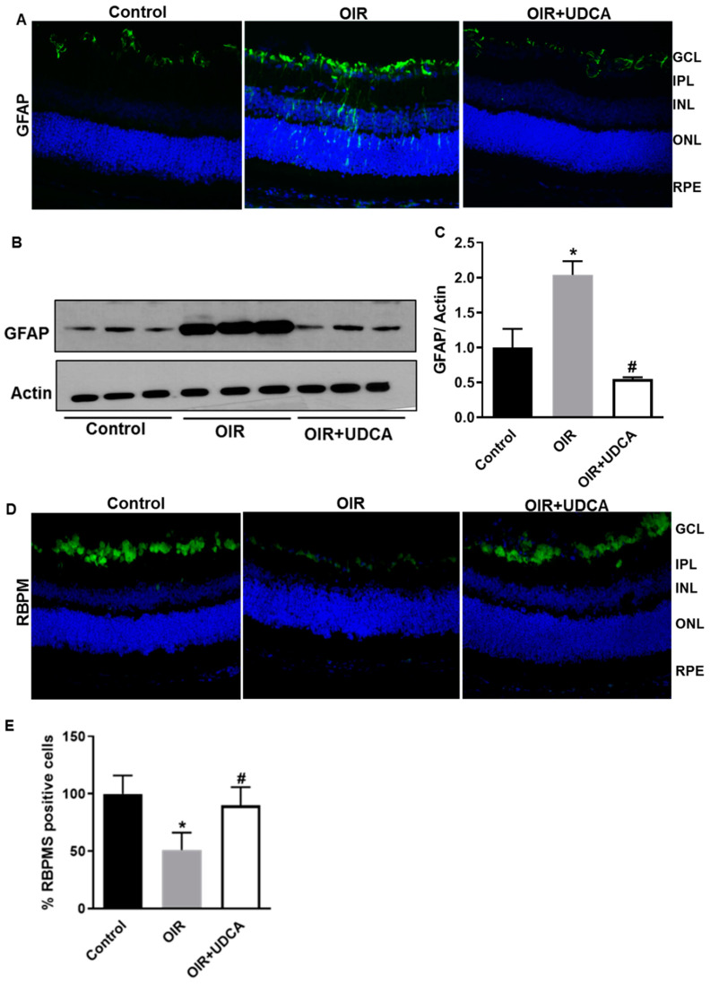
UDCA treatment halts reactive gliosis and preserves neuronal cells in OIR mice. Frozen retinal sections from control mice at P17 (room air), OIR mice, and OIR mice treated with 50 mg/kg UDCA (P7–P17) were stained with (A) GFAP antibody. Total protein fractions from control mice (room air), OIR mice at P17, and OIR mice treated with 50 mg/kg UDCA (P7–P17) were used to analyze (B) protein levels of GFAP by Western blotting. (C) Densitometry graphs showing changes in glial fibrillary acidic protein (GFAP) protein expressions. Values are mean ± S.D. (n = 6 retinas per group). (D,E) Frozen retinal sections from control mice at P17 (room air), OIR mice, and OIR mice treated with 50 mg/kg UDCA (P7–P17) were stained with RNA binding protein with multiple splicing (RBPMS) antibody and positive cells were counted. RBPMS data are expressed as cell number/100 μm retinal length and presented as a percent change from control. * p < 0.05 vs. control and # p < 0.05 vs. OIR. GLC: ganglion cell layer; IPL: inner plexiform layer; INL: inner nuclear layer; ONL: outer nuclear layer; RPE: retinal pigmented epithelium; RBPMS: RNA Binding Protein with Multiple Splicing; GFAP: Glial fibrillary acidic protein.

