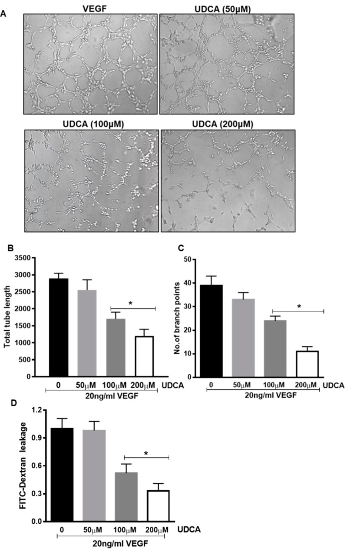
Figure 8.
Effect of UDCA on in vitro tube formation and barrier function in VEGF-treated human retinal microvascular endothelial cells (HuREC). HuREC were maintained on matrigel-coated plates in the presence of 20 ng/mL VEGF alone or in combination with UDCA (50–200 μM). (A) Micro images of tube formation assay. (B,C) Graphs representing results of (B) tube length and (C) number of branch points. Values are mean ± S.D (n = 3). (D) Measurements of FITC (fluorescein isothiocyanate) fluorescence from HuREC grown in trans-well plates after treatment with 20 ng/mL VEGF in presence of different UDCA doses (50–200 μM). * p < 0.05 vs. cells not treated with UDCA.

