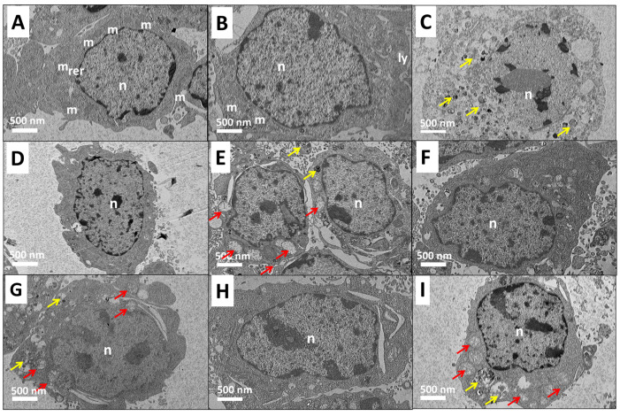
Figure 2.
High-resolution transmission electron microscopy (HRTEM) micrographs of 3T3 mouse fibroblast cells of control (A) at 0 h, (B) at 48 h, and treated groups of 1 mg/mL active ingredient of (C) late apoptosis of dazomet (D) healthy cell of hexaconazole, (E) early apoptosis of hexaconazole, (F) healthy cells of CHEN, (G) early apoptosis of CHEN, (H) healthy cells of CDEN, and (I) early apoptosis of CDEN, at 48 h exposure, where n is a nucleus, m is mitochondria, ly is lysosome, and rer is rough endoplasmic reticulum. The red arrow indicates swollen mitochondria and the yellow arrow indicates condensed vacuoles. Magnification 1500×.

