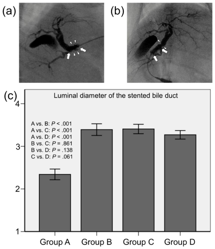
Figure 2.
Representative cholangiographic images showing that SEMSs are fully expanded without stent migration. (a) Follow-up cholangiography in group A shows filling defects (arrowheads) in the stent lumen (arrows) caused by granulation tissue formation and biliary sludge. (b) Follow-up cholangiography showing relatively good patency of the stent (arrows) with no definite irregularities in group B. However, luminal narrowing (arrowheads) at the proximal end of the stent is observed. (c) Mean luminal diameter of the stented extrahepatic bile duct. Note: CI, confidence interval.

