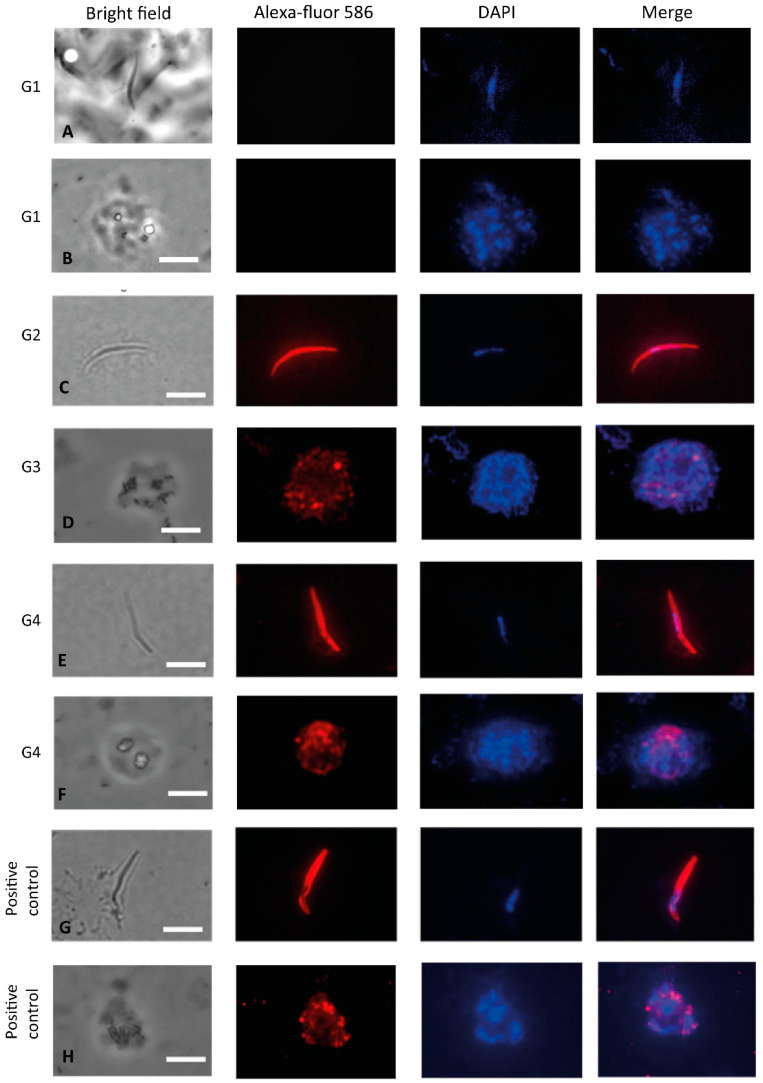
Figure 6.
Indirect immunofluorescence analysis using sera from C57BL/6 mice. Microscope slides containing fixed sporozoites of P. vivax obtained from patients from Thailand (A,C,E,G) and merozoites of P. vivax obtained from patients from Rondônia (Brazil) (B,D,F,H) were incubated with a pool of sera from mice immunized with PvCSP-AllFL, PvAMA-1 or protein mixture in the presence of poly(I:C). (A,B) G1: negative control sera from PBS + poly(I:C), (C) G2: polyclonal sera anti-PvCSP-AllFL (1:100), (D) G3: polyclonal sera anti-PvAMA-1 (1:100), (E) G4: polyclonal sera anti-Mix (1:100), (F) G4: polyclonal sera anti-Mix (1:100), (G) positive control mAb anti-CSP-VK210 (1:100), and (H) positive control mAb K243 anti-PvAMA-1 (1:1000). The white bars are equivalent to 10 µm.

