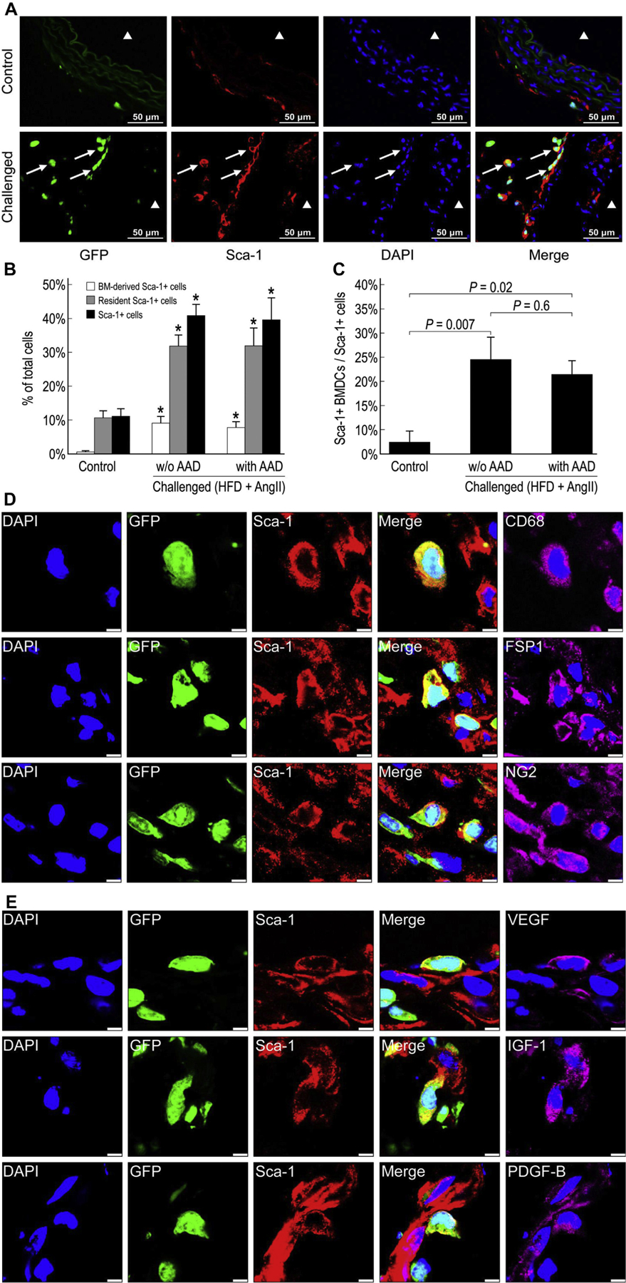
Fig. 8 –

Activation of bone marrow (BM)-derived and resident Sca-1+ stem/progenitor cells in the injured aortic wall. Representative images showing BM-derived Sca-1+ cells in the suprarenal aorta of control and challenged mice (A). ▲ indicates the aortic lumen. White arrows indicate BM-derived Sca-1+ cells. The percentage of BM-derived Sca-1+ cells, resident Sca-1+ cells, and Sca-1+ cells in the total cell population of the suprarenal aorta (*P <0.05 compared with control group) (B) and the percentage of BM-derived Sca-1+ cells in the total Sca-1+ cell population of the suprarenal aorta in challenged and control mice (n = 4 per group) (C). Immunofluorescence staining showing both BM-derived (GFP+) and resident (GFP−) Sca-1+ cells expressing CD68, FSP-1, or NG2 in the aortic adventitia of challenged mice (scale bar = 5 μm) (D). Both BM-derived (GFP+) and resident (GFP−) Sca-1+ cells expressing VEGF, IGF-1, and PDGF-B in the aortic adventitia of challenged mice (scale bar = 5 μm) (E). HFD, high-fat diet.
