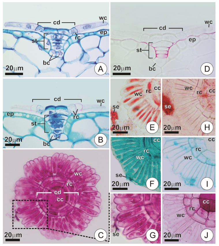
Figure 2.
Anatomy and histochemistry of secretory trichomes. (A,B) Longitudinal section of a trichome showing the overall structure in young (A) and mature (B) bracts. Note the distinct appearance of the wing cells in each of the phases. (C) Shield of a secreting trichome. Note the overall structure and wing cells with conspicuous protoplasts marked for mucilage (in pink). (D) Longitudinal section of a trichome showing cuticle arrangement (in red). (E–J) Portions of the shield showing histochemical characterization of trichomes and secretion in young (E–G) and mature (H–J) bracts. (E,H) Xylidine pounceau, for proteins (in red). (F,I) Alcian blue, for mucilages (in blue). (G,J) Ruthenium red, for mucilages (in pink). (bc = basal cells; cc = central cells; cd = central disc; ep = ordinary epidermis cells; rc = ring cells; se = secretion; st = stalk; wc = wing cells).

