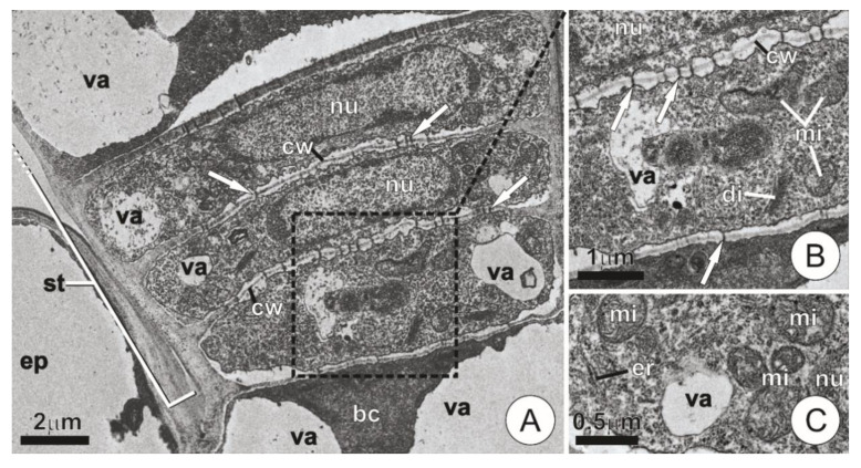
Figure 3.
Ultrastructure of a trichome stalk and basal cells. (A,B) Longitudinal section showing overall aspect (A) and detail of the dotted area (B). Note the cells with thin walls largely connected by plasmodesmata (arrows). (C) Detail of a stalk cell showing dense, mitochondria-rich cytoplasm. (bc = basal cell; di = dictyosomes; ep = ordinary epidermis cell; er = endoplasmic reticulum; mi = mitochondria; nu = nucleus; st = stalk; va = vacuole).

