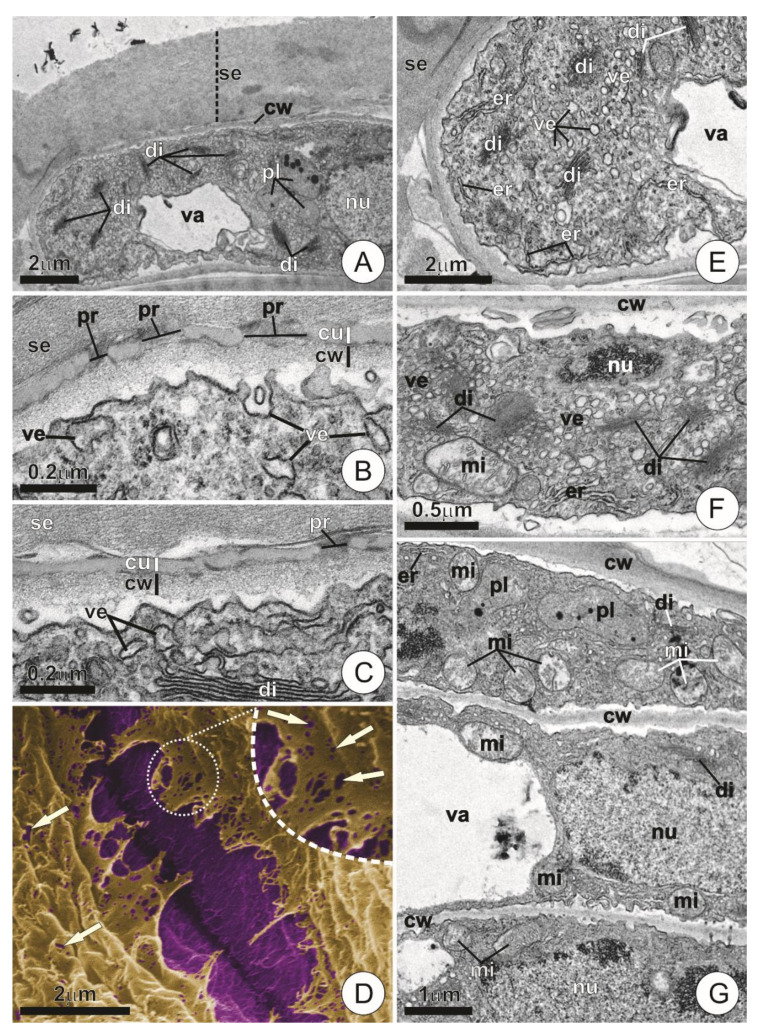
Figure 4.
Ultrastructure of wing cells in young trichomes. (A) Longitudinal section of a wing cell. Note the dense cytoplasm and the thick layer of secretion on the cell surface. (B,C) Detail of the cell wall and protoplast. Note the presence of pores in the cuticle and small vesicles in contact with the plasmalemma. (D) Scanning electron micrograph showing the contact region between adjacent wing cells. Artificial coloring highlights the cell wall (purple) and the cuticle (ruptured, in yellow). Note the presence of cuticular pores (arrows). (E–G) Overview of wing cell protoplasts. Note the dense cytoplasm with numerous vesicles, dictyosomes, and mitochondria. (cu = cuticle; cw = cell wall; di = dictyosome; er = endoplasmic reticulum; mi = mitochondria; nu = nucleus; pl = plastids; pr = pore; se = secretion; va = vacuole; ve = vesicle.).

