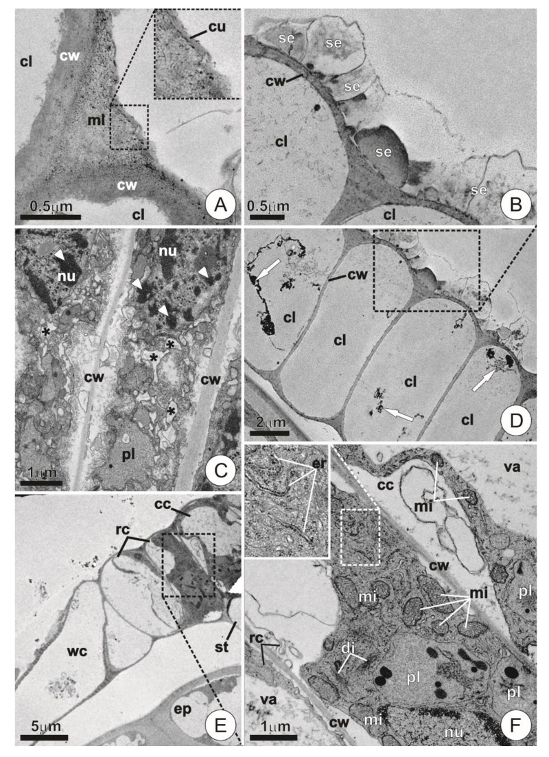
Figure 5.
Ultrastructure of the wing and central disc cells in mature trichomes. (A) Detail of the region between adjacent cells showing conspicuous walls. Note the indistinct and discontinuous cuticle. (B) Detail showing secretion of fibrillar and compact aspect on the surface of mature wing cells. (C) Degenerating wing cells showing chromatin condensation/fragmentation (arrowheads) and the occurrence of vesiculation throughout the cytoplasm (asterisks). (D) Cross-section of the wing showing cells with empty lumens and residues of cytoplasm (arrows). (E) Overview of the shield in cross-section. Note the extravacuolar cytoplasm restricted to the periphery of the outer ring and central cells. (F) Detail of the dotted area in E showing protoplast composition. Note abundant mitochondria, plastids, and endoplasmic reticulum. (cc = central cell; cu = cuticle; cl = cell lumem; cw = cell wall; di = dictyosomes; ep = ordinary epidermis cell; er = endoplasmic reticulum; mi = mitochondria; ml = middle lamella; nu = nucleus; pl = plastids; rc = ring cell; se=secretion; st = stalk; va = vacuole; wc = wing cell.).

