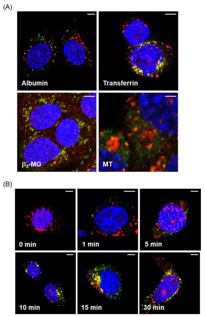
Figure 1.
Fluorescence imaging of endocytosed proteins in mouse proximal tubule epithelial cell (PTEC)-derived S1 cells. (A) S1 cells were incubated with 50 µg/mL fluorescein isothiocyanate (FITC)-albumin, Alexa-transferrin, FITC-β2-MG, or FITC-MT (green) for 30 min and then fixed with paraformaldehyde for immunofluorescence labeling with anti-EEA1 (red). Yellow staining demonstrates the colocalization of fluorescent-labeled proteins and early endosomes. (B) S1 cells were incubated with Alexa-transferrin for 1, 5, 10, 15, and 30 min. The localization of Alexa-transferrin (green) and early endosomes stained with anti-EEA1 (red) was visualized by confocal microscopy. Bars, 5 µm.

