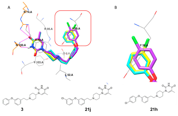
Figure 4.
(A) Structure overlay of compound 3 (cyan), 21j (yellow) and 21h (purple) in the MtbTMPK (PDB 5NR7) [14] active site. (B) Detailed representation of the interactions of the D rings of 3, 21h and 21j, with the side chain of Tyr39. All residues interacting with the inhibitors, including the hydrophobic contact (gray wire) and hydrogen-bonding interaction (residues in orange wire, hydrogen bonds indicated in magenta), were calculated using LigPlus [31]. Illustration was created using Chimera [32].

