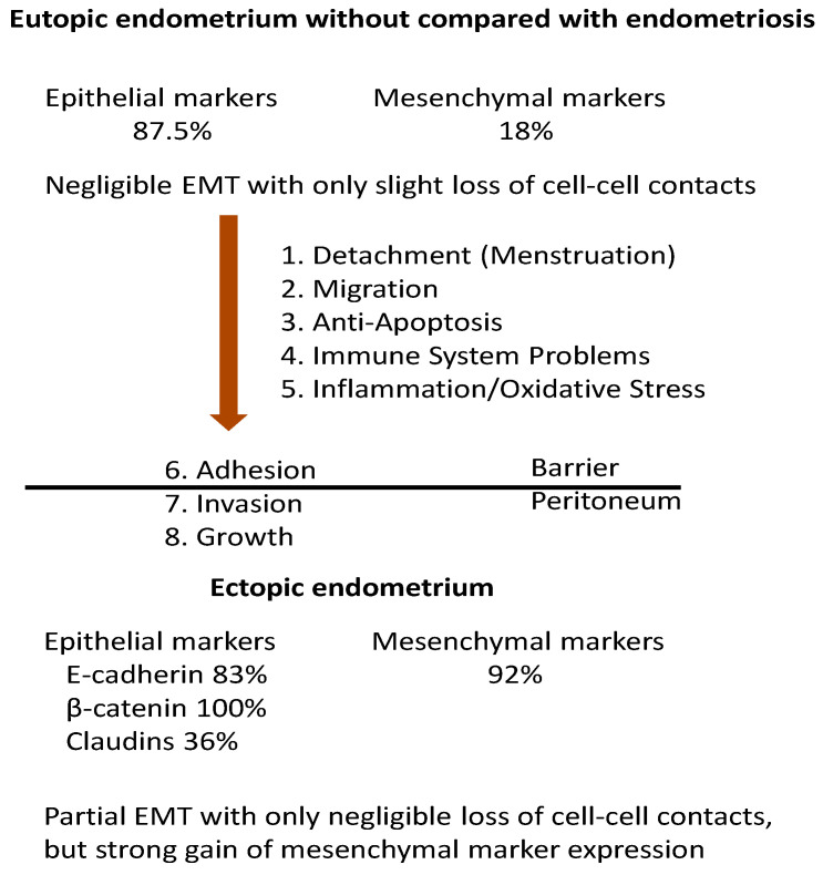
Figure 2.
EMT in endometriosis. At the level of the endometrium, the epithelial EMT markers showed no differences in 87.5% of the studies, whereas the mesenchymal markers showed differences only in 18% of the studies when the eutopic endometrium of patients with and without endometriosis was compared. This suggests a partial EMT with only negligible loss of cell–cell contacts. A decreased protein expression of E-cadherin was found in 83.3% of the studies, but differences in the eutopic endometrium and the ectopic endometrium could be shown in 36.4% of the studies with claudins. The mesenchymal markers in the ectopic endometrium were different in 92% of the studies compared to the eutopic endometrium. Thus, we suggest that, after implantation, EMT was still partial with only a negligible loss of cell–cell contacts but with a strong gain in the expression of mesenchymal markers. In summary, EMT in endometriosis occurs mainly after implantation.

