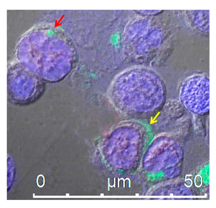
Figure 2.
Confocal microscope analysis of HEK293T cells transfected with an expression vector encoding Nefmut fused to an scFv, labeled with an anti-Nef monoclonal antibody and Alexa 488-conjugated anti-mouse IgG antibody. The picture shows intracellular localization of the fusion protein product in intracytoplasmic vesicle aggregates (red arrow) and at the plasma membrane (yellow arrow). DAPI (blue fluorescence) was used to highlight cell nuclei. Scale bar is shown.

