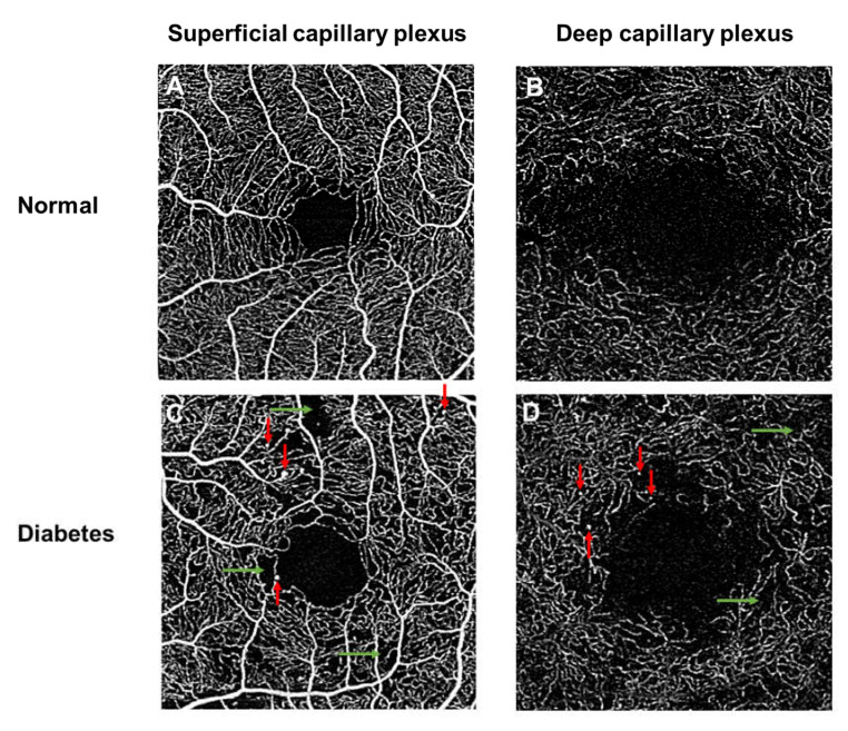
Figure 4.
Optical coherence tomography angiography (OCTA; 3 × 3 mm area) of a healthy control individual (Top panel; A,B), showing an extensive network of capillaries of the superficial vascular plexus, where the foveal avascular zone is surrounded by the foveal capillary network. OCTA images of a patient with diabetes (Bottom panel; C,D), showing vascular abnormalities in both superficial and deep plexus layers, such as microaneurysms (red arrows), capillary nonperfusion (green arrows). Note the enlarged foveal avascular zone (FAZ). (B,D) were created after removal of projection artifacts.

