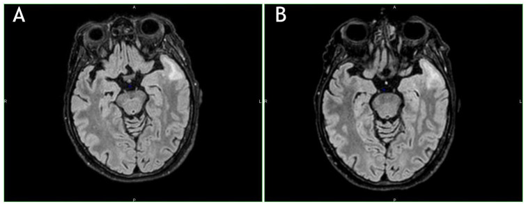
Figure 2.
(A) Hyperintense lesions in the left frontotemporal pole along with confluent juxta-cortical lesions of the white matter of the left temporal pole and homolateral insula. T2-FLAIR weighted image. (B) Further new magnetic resonance imaging performed 3 weeks later. Note the slight dimensional increase of the confluent temporal lesion. T2-FLAIR weighted image.

