Figure 3.
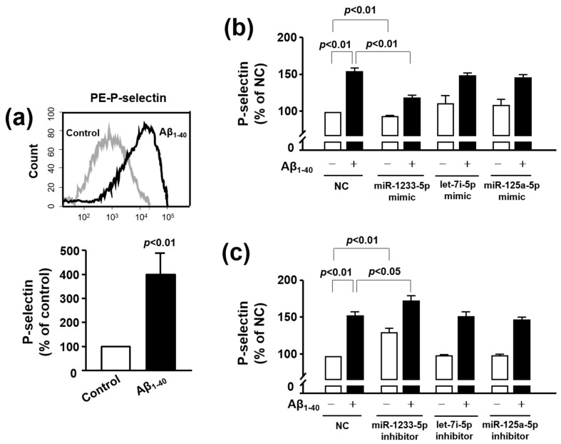
Role of let-7i-5p, miR-125a-5p, and miR-1233-5p in the Aβ1-40-induced P-selectin expression in platelets and MEG-01 cells. (a) Platelet activation status at basal level (gray curve) and after 10 μM Aβ1-40 stimulation (black curve) was monitored by flow cytometric measurement of PE-P-selectin expression. Data are mean ± SEM of six experiments. (b and c) Overexpression or inhibition of let-7i-5p, miR-125a-5p, and miR-1233-5p expression resulted in the alteration in the level of P-selectin in MEG-01 cells. Let-7i-5p, miR-125a-5p, and miR-1233-5p were individually overexpressed (b) or downregulated (c) using a specific mimic or inhibitor in MEG-01 cells after treatment with or without 10 μM Aβ1-40 for 1 h. P-selectin level was modulated in these samples as compared to that in samples treated with MEG-01 negative control (NC). Data are mean ± SEM of six experiments.

