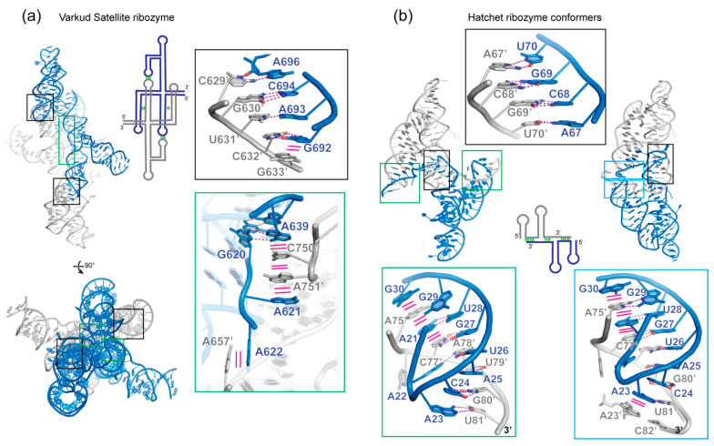
Figure 3.
Structures of dimeric ribozymes. (a) Crystal structure of Varkud Satellite ribozyme (PDB code 4R4P & 4R4V) with the insets showing the intermolecular kissing loop interaction between stem-loop 1 (P1) and stem-loop 5′ (P5′), as well as the intercalation of stacked nucleobases between P1 and P6′. (b) Crystal structures of the Hatchet ribozyme (PDB 6QJ6 & 6QJ5) showing two types of dimeric interfaces. The top inset highlights the palindromic sequence paired with the same sequence of the second protomer. Hydrogen bonding interactions are shown as magenta dashed lines, while stacking interactions are shown as solid magenta lines.

