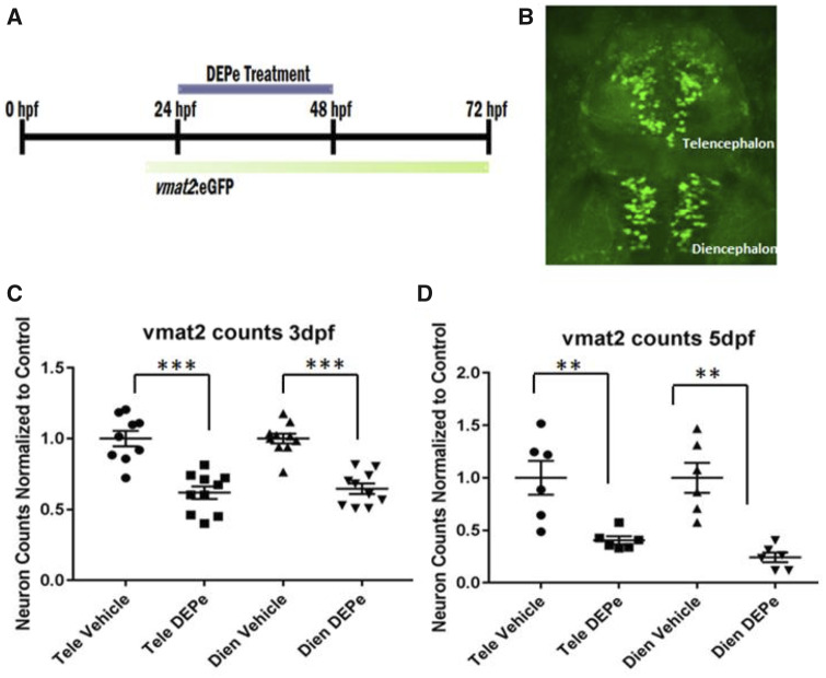
Figure 2.
DEPe treatment and analysis of ZF aminergic neurons. Experimental design (A) and representative image of the aminergic neurons quantified in the vmat2: eGFP transgenic line (B). Vmat2+ neurons were quantified after treatment with 10 µg/ml DEPe at (C) 3 dpf or (D) 5 dpf via confocal microscopy in fixed embryos and counts were normalized to the baseline in control embryos for aminergic neurons in the telencephalon (Tele) and diencephalon (Dien). Counts from 3 and 5 dpf showed a statistically significant decrease in neurons after DEPe treatment (***p < .0001, **p < .005 as determined by one-way ANOVA with Sidak’s multiple comparisons analysis and SEM).

