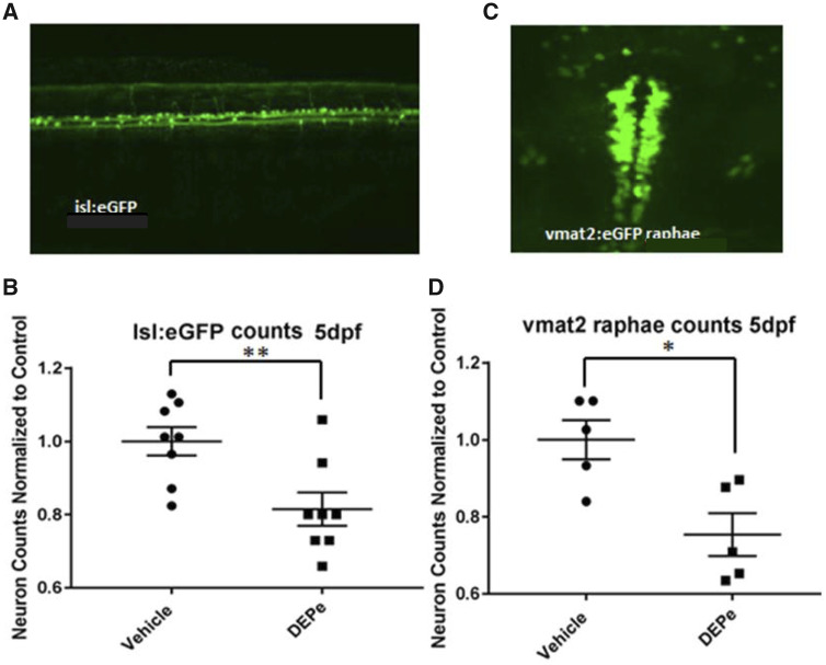
Figure 3.
Specificity of neuronal toxicity after DEPe exposure. Neurons expressing GFP from the islet1 or vmat2 promoter were quantified at 5 dpf after 24 h of DEPe treatment in order to determine the selectivity of neurotoxicity (A). We found a significant loss of GFP-positive sensory neurons in the tail of isl:GFP embryos after treatment with 25 µg/ml DEPe (B). Statistical analysis by unpaired two-tailed Student’s t test (**p = .0077) (C). Serotonergic neurons within the raphae cluster of vmat2: eGFP-positive embryos treated with 10 µg/ml DEPe were also significantly reduced at 3 dpf as quantified by unpaired two-tailed Student’s t test (*p = .011) (D).

