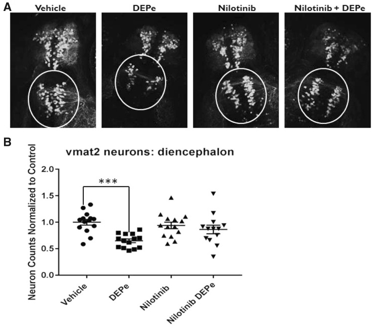
Figure 6.
Testing neuroprotection with nilotinib after DEPe treatment. Vmat2 positive neuron counts from blinded confocal images after treatment with DEPe and/or nilotinib (A). Data represent diencephalic neuron counts from 3 experiments, n = 14 larvae normalized to control counts for each experiment. DEPe alone shows a significant reduction by one-way ANOVA with Dunnett’s multiple comparison analysis (***p = .0003) in the number of aminergic neurons in the diecephalon (B). Neither nilotinib alone nor the combined nilotinib/DEPe treatments show a significant change in neuron number relative to control.

