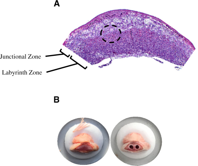
Figure 1.

A, A representative hematoxylin and eosin stained placental section depicting location of the junctional and labyrinth zones. The dotted circle indicates the region where the micropunches were obtained. B, Representative images of a placenta mounted on the cryostat before and after two 1.25-mm micropunches were collected.
